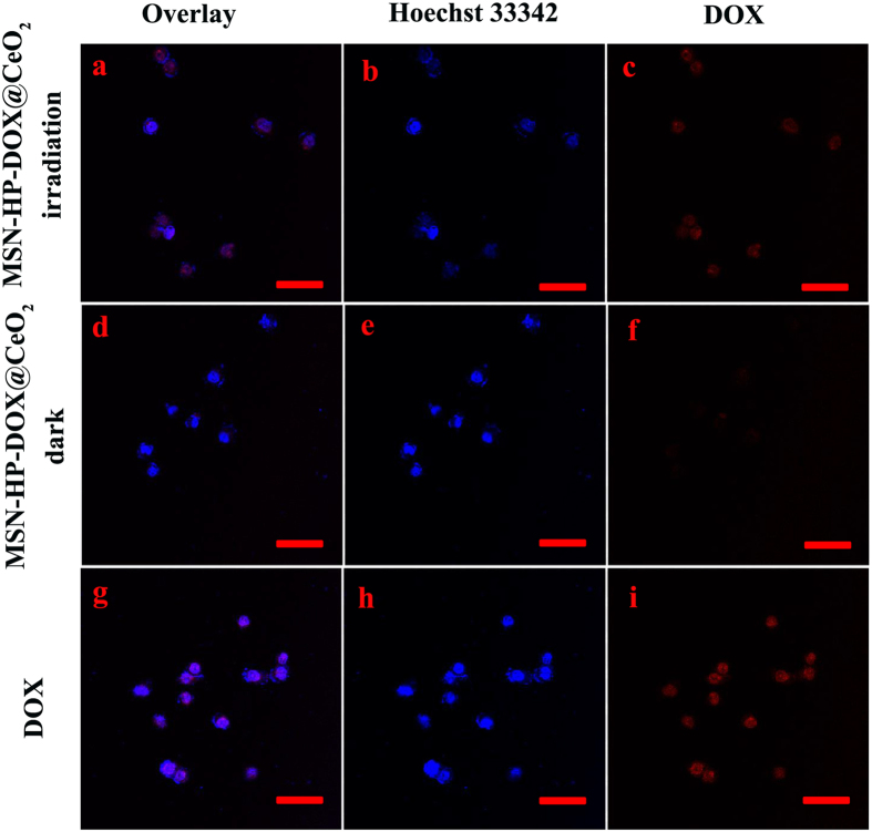
Figure 6.
CLSM images of HeLa cells incubated with MSN-HP-DOX@CeO2 accompanied with light irradiation (a–c), MSN-HP-DOX@CeO2 without light irradiation (d–f) and DOX (g–h): panels b, e and h are blue channels with Hoechst 33342; panel c, f and i are red channels for DOX; panels a, d and g are merged images of panels b,c, e-f and h,i, respectively. Scale bar: 50 μm.

