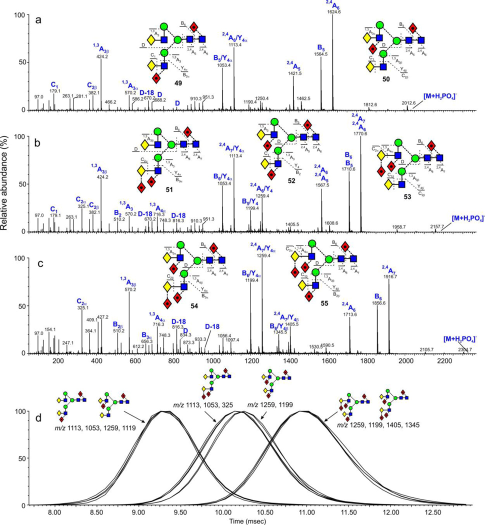
Figure 11.
(a) Negative ion CID spectra of the di-fucosylated biantennary N-glycans from parotid glycoproteins. (b) Negative ion CID spectra of the tri-fucosylated biantennary N-glycans from parotid glycoproteins. (c) Negative ion CID spectra of the tetra-fucosylated bantennary N-glycans from parotid glycoproteins. (d) Extracted fragment ATDs of diagnostic ions (see text) defining the isomers of the tri-fucosylated biantennary glycans. Fragment ATDs were smoothed with the mean algorithm (window size ± 2 scans) from MassLynx and are all normalized to 100%.

