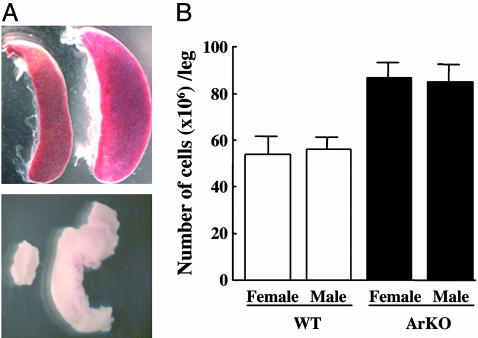
Fig. 1.
Enlarged spleen, mesenteric lymph nodes, and bone marrow hyperplasia in ArKO mice. (A) Photograph of spleen (Upper) and mesenteric lymph nodes (Lower) of WT (left) and ArKO (right) mice. Pictures are representative of at least six mice killed for each sex and genotype. (B) Twelve- to 16-month-old ArKO (black columns) and age-matched WT (white columns) male and female mice were killed and, from each mouse, one femur and one tibia were flushed with PBS. The cells were washed and resuspended in 10 ml of PBS. The total cells per leg were calculated based on hemocytometer counts (data are shown as mean; n = 6 per group).

