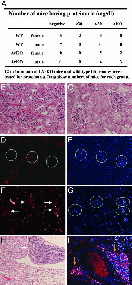
Fig. 3.
Presence of proteinuria and infiltrating leukocytes in the perirenal area of the kidney from ArKO mice. (A) Impaired renal function was confirmed by the presence of significant proteinuria in ArKO mice. (B, C, and H) Kidney sections from old WT (B) and ArKO (C and H) mice stained with hematoxylin/eosin. (D–G) Deposition of IgG was detected by immunofluorescence using kidney sections from WT (D and E) and ArKO (F and G) mice. Nuclei counter-stained with 4′,6-diamidino-2-phenylindole are shown in E and G. White circles, location of glomeruli; white arrows, IgG deposition in the glomeruli. (H and I) There is a severe infiltration of B lymphocytes in kidneys from ArKO mice (white arrow, area of infiltration; yellow arrows, plasma B cell-related phenotype, showing intracellular Ig), whereas WT littermates are normal.

