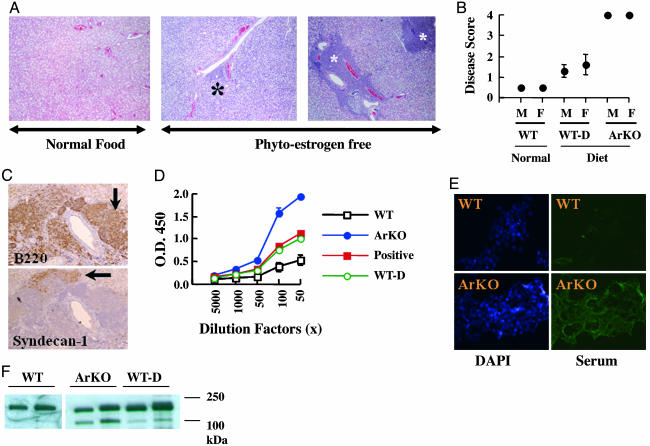
Fig. 4.
ArKO mice develop a SS-like phenotype as they age. (A) Paraffin sections of salivary glands from WT mice fed a normal diet (Left), WT mice fed a phytoestrogen-free diet (Center, WT-D), and ArKO mice fed a phytoestrogen-free diet (Right) were stained with hematoxylin/eosin. Asterisks, periductal infiltrates (foci). (B) Paraffin sections of salivary glands from six mice from each group were prepared as shown in A and scored for disease as described in Materials and Methods. (C) B220+ B lymphocytes are dominant in the lymphoepithelial lesions in ArKO mice, and some of these lymphocytes are characterized as plasma B cells showing syndecan-1 positivity. (D) Anti-α-fodrin antibodies were detected in sera from WT and ArKO mice by ELISA. (E) Sera from ArKO mice show immunoreactivity against cytoskeletal proteins in cultured MCF-7 cells. DAPI, 4′,6-diamidino-2-phenylindole. (F) Detection of α-fodrin cleavage products in salivary gland from WT (lanes 1 and 2, from left to right), ArKO (lanes 3 and 4), and WT-D (lanes 5 and 6) mice.

