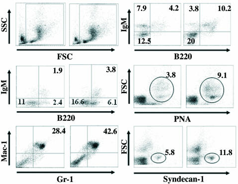
Fig. 5.
Increased number of B lymphocytes in ArKO mice. (Top Left) Side scatter/forward light scatter plots of bone marrow cells from ArKO (Right) and WT (Left) mice are shown. Leukocytes were gated, as shown in the plots for each analysis. (Middle Left) Increased B220+ B cells from bone marrow of ArKO mice. (Bottom Left) Increased Gr-1+/Mac-1+ myeloid cells from bone marrow of ArKO mice. (Top Right) Freshly prepared splenocytes were stained with antibody against IgM and B220 and significant increase in B220+ B cells was observed, whereas the percentage of IgM+/B220– B cells was decreased in ArKO mice. Increased percentages of peanut agglutinin-positive germinal center B cells (Middle Right) and plasma B cells (syndecan-1 positive) (Bottom Right) are shown. Percentages of each cell type from a total of 106 cells are indicated in each box. The figures are representative of five different female mice.

