Fig. 8.

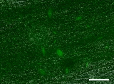
Light microscopy image of lack of neurite extension on PCL fibers. Cells are stained green with beta-III-tubulin. Scale bar is 100 μm

Light microscopy image of lack of neurite extension on PCL fibers. Cells are stained green with beta-III-tubulin. Scale bar is 100 μm