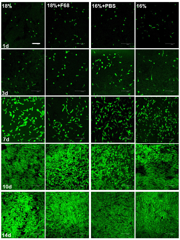
Figure 2.

Confocal micrographs of MG63 cells in scaffolds stained with FDA after different cultivated times. The confocal images were taken along z-sections into the scaffolds and shown as 2D pictures composed by Leica Confocal Software. Scale bar, 150 μm.
