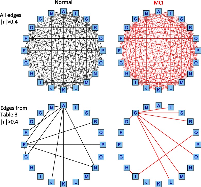
Fig. 2.

Correlation networks for the 20 miRNAs detected by differential correlation analysis. Each box indicates a miRNA with the alphabet in Table 5. For example, A: hsa-miR-191, B: hsa-miR-590-5p, C: hsa-miR-125b, D: hsa-miR-18a, E: hsa-miR-140-3p and F: hsa-miR-103. The 10 miRNAs (A, B, E, F, G, J, K, N, P, R) and the 11 miRNAs (C, D, H, I, L, M, N, O, Q, R, S, T) are highly correlated with each other in Normal and MCI, respectively. Upper: all edges with the correlation coefficient of |r|>0.40. Lower: the edges with the correlation coefficient of |r|>0.40 only for differentially correlated miRNA pairs in Table 3. Solid and broken lines indicate positive and negative correlations, respectively
