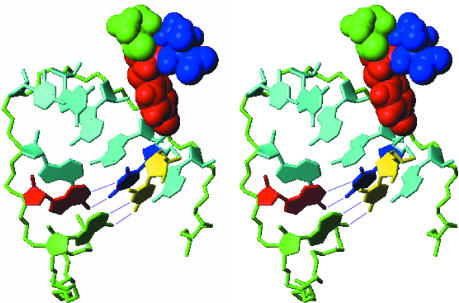
Fig. 5.
Stereoview of a model showing potential base-pairing between pUpU and the critical adenosine residues of the cre that template uridylylation. The middle segment of the loop is shown with the A15 (green) and A16 (red) residues forming conventional Watson-Crick base pairs with pUpU (yellow and blue). The polypeptide component of VPg is coupled to the uppermost uridine residue (blue) in this view. The tyrosine residue of VPg (red) is shown, with the flanking residues, Ala (green) and Thr (blue), also shown in the model. The phosphate backbone of the cre loop is green, and residues A17-A22 are cyan.

