
Fig. 2.
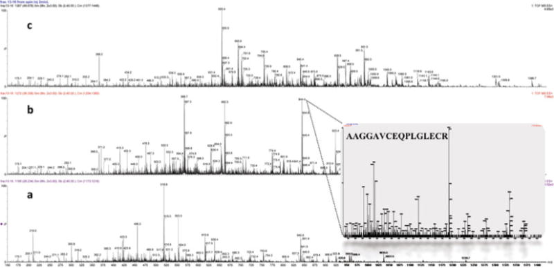
Mass spectra of the MUC5B digest over different retention time scans during the capillary reverse-phase separation. Typical survey scan mass spectra of: (a) early (22–28 min), (b) middle (30–40 min), and (c) late (42–50 min) eluted peptides. Inset spectra: MS/MS spectra of the doubly charged precursor ion 844.41 Da, AAGGAVCEQPLGLECR.