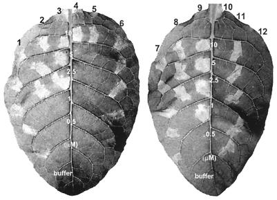
FIG. 3.
Comparison of the HR elicitor activity in tobacco leaves of HpaG mutant proteins, the HpaG peptide, and the HpaG(L50P) peptide. The proteins were infiltrated into tobacco leaves at concentrations of 10, 5, 2.5, 1, or 0.5 μM in 20 mM Tris-HCl (pH 8.0). Labeling: 1 and 7, HpaG; 2, HpaG(L39A); 3, HpaG(L39P); 4, HpaG(L42D); 5, HpaG(L43P); 6, HpaG(Q45A); 8, HpaG(L46A); 9, HpaG(L50A); 10, HpaG(L50P); 11, HpaG peptide; 12, HpaG(L50P) peptide; buffer, 20 mM Tris-HCl (pH 8.0). Tobacco (N. tabacum cv. Samsun NN) leaves were photographed 24 h after infiltration.

