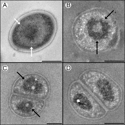
FIG. 1.
Transmission electron micrographs of cryofixed D. radiopugnans (A and C) and D. radiophilus (B and D) cells. (A) Regular staining. The darkly stained particles are ribosomes, while the lightly stained space contains chromatin. (B, C, and D) Cells stained with the DNA-specific reagent osmium-ammine-SO2 (27). DNA toroids (indicated by arrows) are evident in panels A, B, and C, whereas in panel D the toroids are detected edge on. Because thin sections are used, some (cross-sectioned) specimens reveal only one compartment. Scale bars, 0.5 μm.

