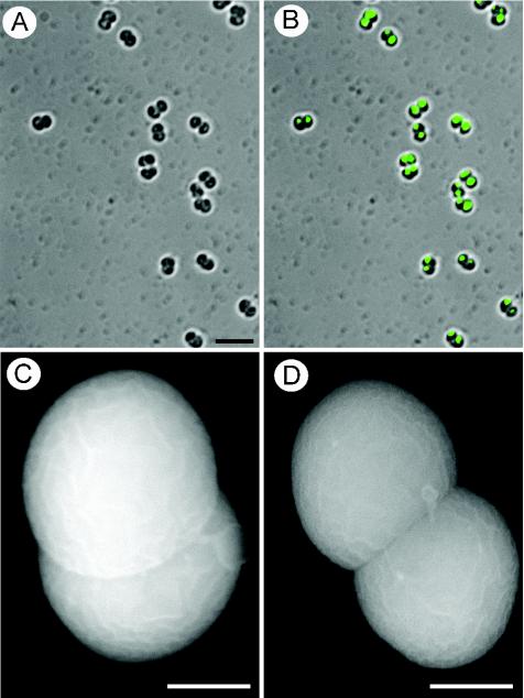
FIG. 2.
Morphology and DNA segregation in D. radiopugnans cells from 4-day-old cultures. (A and B) Shown are light (A) and fluorescence (B) microscopy of cells labeled with DAPI (4′,6′-diamidino-2-phenylindole). DNA segregation in both compartments of each diplococcal unit is evident. A diplococcal morphology is demonstrated by all cells, as indicated by both light (A and B) and scanning electron (C and D) microscopy. Scale bars, 5 (A and B) and 0.5 (C and D) μm.

