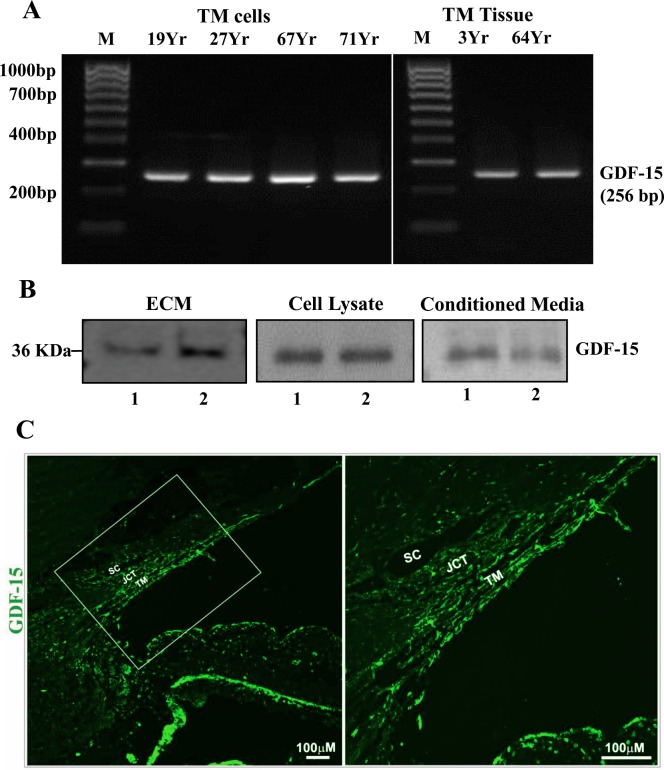
Figure 1.
Expression and distribution of GDF-15 in human TM cells, tissue, and the AH outflow pathway. (A) Reverse transcription-PCR based detection of GDF-15 expression in TM cells derived from several individual human donor eyes and TM tissue. (B) Immunoblotting-based detection of GDF-15 protein in the SDS-urea soluble ECM fraction, cell lysates, and conditioned media from two independent primary cultures of human TM cells (lanes 1, 2). (C) Immunofluorescence (green)–based analysis of GDF-15 distribution in the human AH outflow pathway. The panel on the right shows a magnified image of the boxed portion in the left panel. Scale bars: Indicate image magnification.

