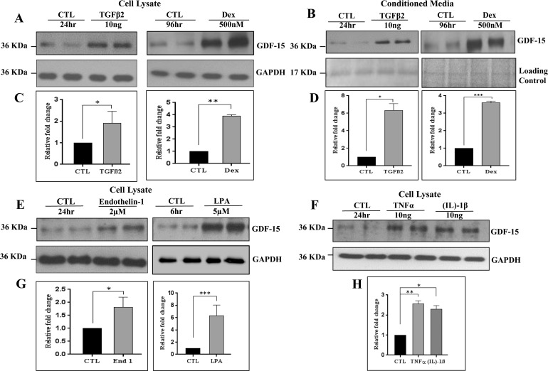
Figure 2.
Regulation of GDF-15 protein levels in human TM cells. (A, C, E–H). Confluent serum-starved TM cells treated with TGFβ2 (10 ng/ml, 24 hours), dexamethasone (500 nM/4 days), endothelin-1 (2 μM/24 hours), LPA (5 μM/6 hours), TNFα (10 ng/ml, 24 hours), and IL-1β (10 ng/ml, 24 hours) showed a significant increase in GDF-15 protein levels in cell lysates compared to control cells (CTL) based on immunoblot analysis. (B, D) Similarly, conditioned media derived from the TGF-β2 and dexamethasone treated TM cells showed a significant increase in GDF-15 protein levels compared to control cells. Histograms depict the fold change in GDF-15 protein levels in the above described samples compared to controls based on densitometric analysis. Glyceraldehyde-3-phosphate dehydrogenase was immunoblotted as a loading control for the cell lysates. Protein loading was normalized for conditioned media samples by subjecting the equal amounts of protein to SDS-PAGE analysis and staining with gel code blue. Staining intensity of the indicated protein band (17 kDa) was used for normalization. Values represent the mean ± SEM of 4 independent determinations. *P ≤ 0.05; **P ≤ 0.01; ***P ≤ 0.001.

