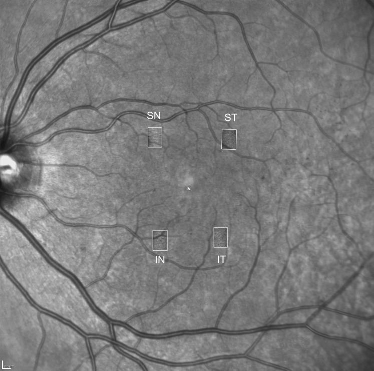
Figure 1.
Overlay of a wide-field infrared image with macular AOSLO images. The white rectangles indicate the location of the four regions of interest for AOSLO imaging: superior-nasal (SN), superior-temporal (ST), inferior-temporal (IT), and inferior-nasal (IN). The asterisk indicates the center of the fovea. Scale bar: 200 μm.

