
Fig 2.
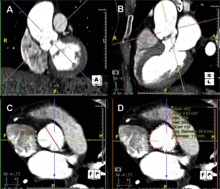
(A) Coronal and (B) sagittal views of the root and ascending thoracic aorta demonstrate representative landmarks: sinotubular junction and anulus. Orthogonal imaging planes show the (C) sinus to commisure diameter (diagonal line) and (D) area (circular line) quantification methods of the level of the sinuses of Valsalva.