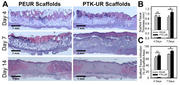
Figure 2.
PEUR and PTK-UR scaffolds implanted into diabetic excisional wounds (A) were largely degraded by 14 days and covered by new epidermis. Implanted PTK-URs promoted (B) greater wound stenting/thicker granulation tissue formation and (C) supported more robust tissue infiltration over 7 days (mean ± SEM, n = 7 independent animals, *p<0.05).

