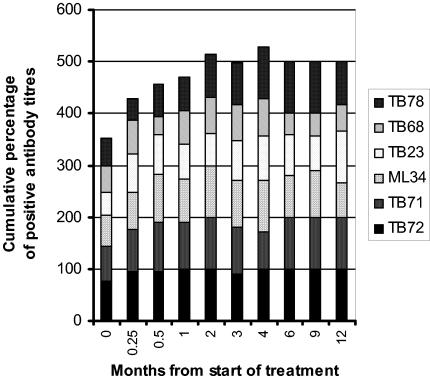
FIG. 2.
Recognition of different epitopes during treatment for tuberculosis. Cumulative percentages of positive titers of antibodies to the six epitopes are plotted against time from the start of treatment. This figure includes sera from patients with smear-positive pulmonary tuberculosis who were ever positive for antibody to the epitopes defined by the monoclonal antibodies TB72 and TB71 (38-kDa secreted antigen), ML34 (lipoarabinomannan), TB23 (19-kDa secreted antigen), TB68 (16-kDa α-crystallin stress protein homologue), and TB78 (stress protein hsp65). Those with isoniazid-resistant or previous tuberculosis have been excluded, as have those with smear-negative pulmonary disease. A positive titer was defined as greater than the mean plus two SD of control sera.

