Fig. 2.
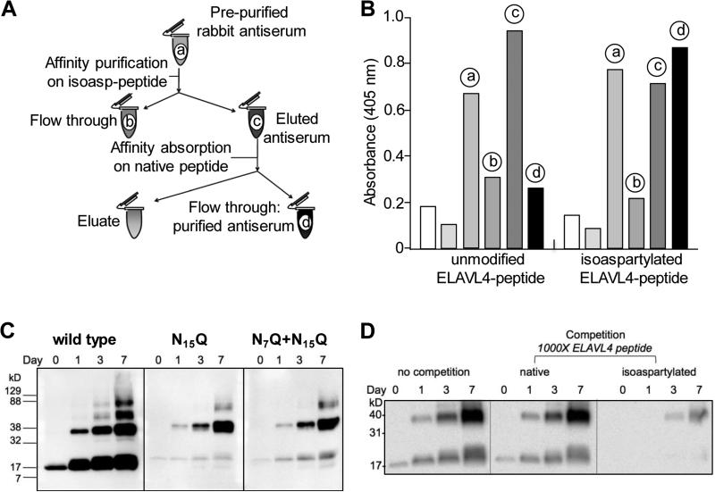
An anti-isoAsp ELAVL4 antiserum detects isoaspartylation in vitro (A) Schematic representation of polyclonal rabbit anti-isoAsp ELAVL4 antiserum purification. The labels a-d match those in the histogram in panel B. (B) ELISA analysis of pre-purified serum (a, 1:100,000 dilution), affinity purification flow-through (b, 1:100,000 dilution), affinity purified eluted antibody (c, 0.01 μg/ml), affinity-absorbed antibody (d, 0.01 μg/ml), and no antibody controls (leftmost white and grey bars) against an unmodified ELAVL4 peptide and the same peptide containing an isoaspartate residue at N15 (see Figure 1B, underlined). ELISA data provided by YenZym Antibodies, South San Francisco, CA. (C) The isoAsp-ELAVL4 antisera specifically recognizes in vitro isoAsp-converted ELAVL41-117. 2 μg wild type, single mutant N15Q and double mutant N7Q+N15Q protein per lane were in vitro isoaspartyl converted at 37°C for up to seven days. Samples from day 0, 1, 3, and 7 were subjected to SDS-PAGE and Western blot analysis and exposed for 5 minutes. A representative example of triplicate experiments is shown. (See Pulido et al. 2016 for reactivity of antiserum with other ELAVL family members.) (D) Competition assay using the same ELAVL4 peptide against which the rabbit antisera had been raised, in its native or isoaspartylated form.

