Fig. 3.
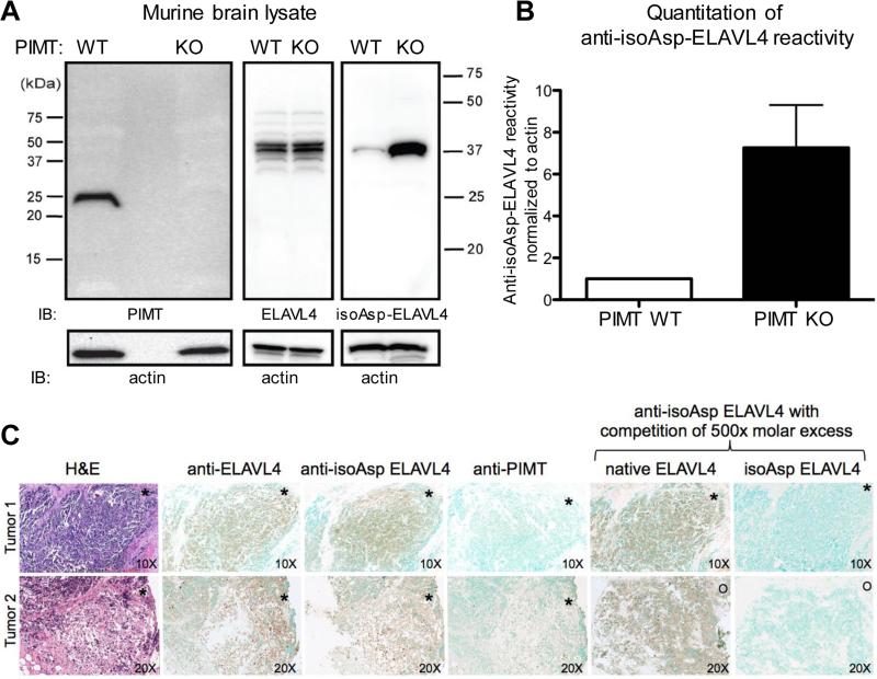
Small cell lung cancer antigen ELAV is prone to isoaspartylation in vivo. (A) Brain extract from PIMT wild type and knock out mice was subjected to Western blot analysis. The brain is the normal site of ELAVL4 expression. Blots were probed with anti-PIMT and anti-ELAVL4 antibodies and the rabbit affinity-purified anti-isoAsp ELAVL4 antiserum. Membranes were stripped and reprobed using an anti-actin antibody to check for similar loading. Exposure time was 45 seconds. (B) Quantitation of isoAsp-ELAVL4 reactivity. (C) Anti-ELAVL4 reactivity of human archival SCLC tumors. Two representative cases are shown out of 5 examined. De-identified archival paraffin blocks were sectioned and subjected to H&E staining or immunohistochemistry with antisera to ELAVL4, isoAsp-ELAVL4, and PIMT. Specificity of the anti-ELAVL4 antiserum was assessed by competition with excess native or isoAsp-converted ELAVL41-117. Asterisks and circles are provided for sample orientation purposes. Top panel, magnification of sections 10X; lower panel, 20x.

