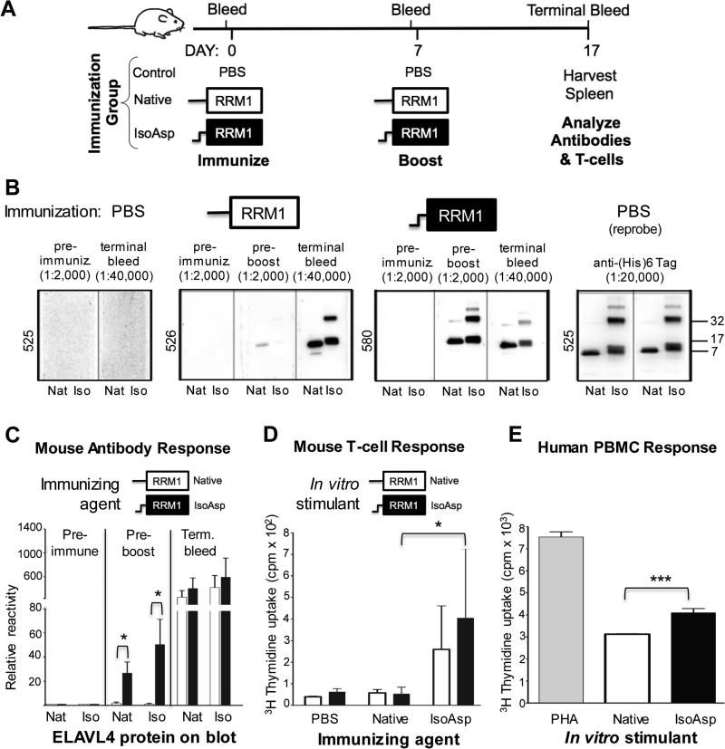
Fig. 4.
Isoaspartylated ELAVL41-117 is highly immunogenic. (A) Schematic depicting strategy to assess the immunogenic potential of isoaspartylated-ELAVL4 in mice. Three mice each, immunized with native ELAVL41-117, the same protein incubated under isoaspartylation conditions for 7 days, or PBS were used. (B) Anti-ELAVL4 and anti-isoAsp-ELAVL4 reactivity of mouse plasma was determined as in Fig. 2C by western blot analysis using 0.25 μg recombinant protein in its native form (Nat) or incubated under isoapsartylation conditions for 7 days (Iso); dilutions of mouse plasma are indicated at top (post-boosting sera were more diluted). The right-most panel shows rehybridization of the strips from mouse 525 with an anti-(His)6 tag antibody. (C) Quantification of mouse antibody response. (D) T-cell proliferation comparing mice immunized with native vs. isoAsp-ELAVL41-117. (E) Induction of human immune cell proliferation in vitro with isoaspartylated ELAVL41-117. Human PBMC from four unidentified donors were incubated in triplicate with native ELAVL41-117, isoAsp-ELAVL41-117 or positive control phytohemagglutinin (PHA) 72 h prior to [3H]thymidine pulse. Total cell proliferation was then measured at 96 h. This graph is a representative example of an experiment. * p<0.05, *** p<0.001.

