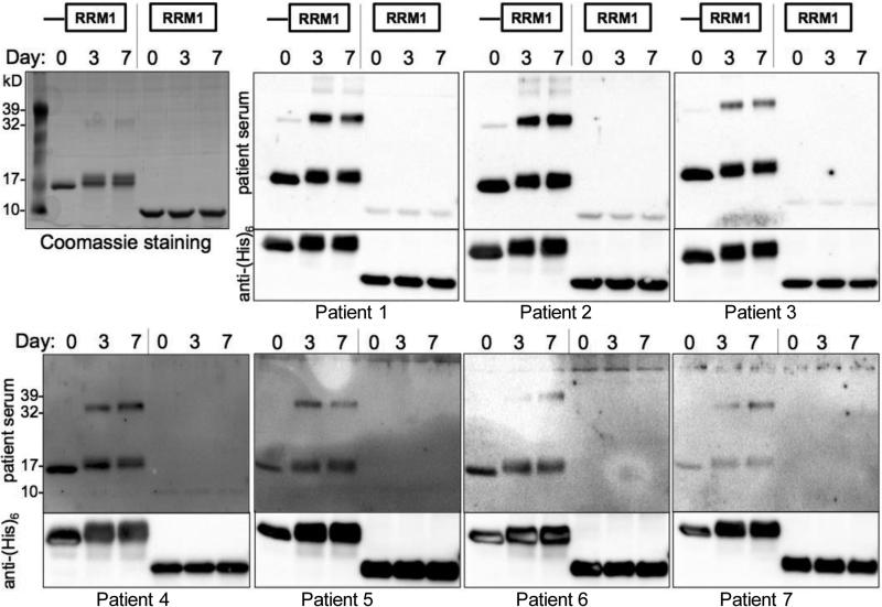
Fig. 5.
SCLC patient sera target the isoAsp-prone region. Recombinant ELAVL41-117 and RRM1 alone were subjected to isoAsp-inducing conditions and gel electrophoresis. The top left panel shows a Coomassie stain of a representative gel. As the protein acquires isoaspartylation (Day 3, 7), mobility changes, widening the band on gel. The other panels show blots probed with patient antiserum (top section) or stripped and reprobed with an anti-(His)6 antibody (1:10,000-20,000) recognizing the C-terminal hexahistidine tag on the recombinant proteins (only bottom section of each panel is shown to indicate protein presence). Top row: 1:10,000 human serum dilution. Bottom row 1:1,000 human serum dilution, except for patient 7, 1:500 dilution. Note the weak or absent staining of the RRM1 alone fragment with the human sera.

