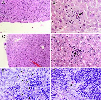
FIG. 2.
Comparison of hepatic pathology (A and C) and localization of A. phagocytophilum-infected cells in livers (B and D) and spleens (E and F) of SCID mice treated with either anti-CXCR2 or control antibody before challenge. Note scattered inflammatory lesions and the presence of scattered infected cells (arrows) in liver and an increased concentration and clustering of infected cells (arrowheads) in the spleens of control antibody-treated SCID mice (E) compared with anti-CXCR2-treated SCID mice (F). These panels show typical examples revealed by these methods in duplicate studies. Tissues from anti-CXCR2-treated (A, B, and F) and control antibody-treated (C, D, and E) animals are shown. Magnifications, ×64 (H&E [A and C]), ×252 (B and D), and ×400 (immunoalkaline phosphatase with rabbit anti-A. phagocytophilum [E and F]).

