
Fig. 4.
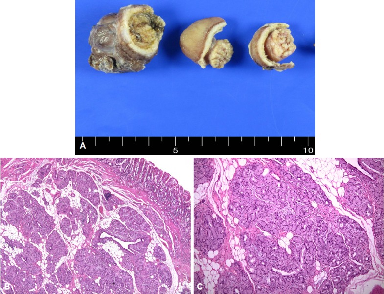
Gross findings after formalin fixation. (A). A well-defined heterogenous yellow-white solid duodenal mass can be observed on the specimen section. The gross endoscopic resection specimen showed a large duodenal lesion measuring 9.3×2 cm. (B) Microscopic findings. Light microscopy revealed hyperplastic lobules of proliferating Brunner's glands separated by fibrous septum. (H&E stain, ×40). (C) Brunner's gland hyperplasia composed of variable size of Brunner’s glands (H&E stain, ×100) can be observed.