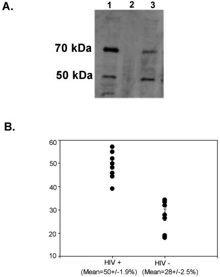
FIG. 2.
(A) Western blot. RTF is expressed in two different forms in HIV-positive and HIV-negative individuals. Lymphocytes from HIV-negative individuals (lane 1) and HIV-positive individuals (lanes 2 and 3) were tested by Western blotting and probed with monoclonal antibody 2C1. Top row, 70-kDa RTF protein; bottom row, 50-kDa RTF protein. The results are representative of those for lymphocytes from HIV-negative individuals and HIV-positive individuals that express RTF (lane 3) and that do not express RTF at detectable levels (lane 2). (B) The percentage of the 50-kDa RTF protein is increased in lymphocytes from HIV-positive individuals. The results are the percentages of the 50-kDa RTF protein expressed by nine HIV-positive individuals and seven HIV-negative individuals, as measured by desitometry. Means are presented with standard errors. The results were deemed to be significant if the P value was <0.001 by Student's t test.

