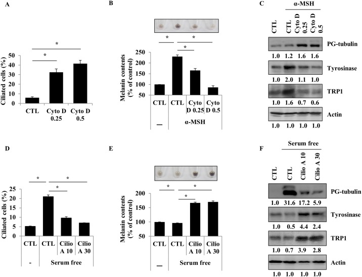
Fig 3. Ciliogenesis negatively regulates melanogenesis in B16F1 cells.
B16F1 cells were treated with cytochalasin D (0.25 or 0.5 μM) for 48 h. The proportion of ciliated cells was measured (A). B16F1 cells were pre-treated with α-MSH (1 μM). After 24 h, they were treated with cytochalasin D (0.25 or 0.5 μM). After 48 h, cellular melanin content was measured (B). Polyglutamylated-tubulin, tyrosinase, and TRP1 were analyzed (C). B16F1 cells were treated with ciliobrevin A1 (10 or 30 μM) in serum-free media for 48 h. The proportion of ciliated cells was evaluated (D) and cellular melanin content was measured (E). Polyglutamylated-tubulin, tyrosinase, and TRP1 were analyzed (F). Data represent the mean ± SE of 3 experiments (* p < 0.05, ** p < 0.01).

