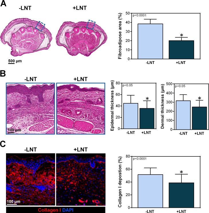
Fig 2. LNT decreases the pathological changes of lymphedema.
A) Left panel: Representative H&E stain of the hindlimbs of mice with or without LNT. Cross-sections were obtained 2 mm proximal to the tarsal joint. The dotted black line indicates the area of fibroadipose deposition. The area highlighted by the blue dotted box is shown in high-power view in part B. Right panel: Quantification of the percentage of fibroadipose deposition area of hindlimbs of mice with and without LNT. B) Left panel: Representative high-power view of the areas indicated in the blue dotted boxes in part A. Note the decreased hyperkeratosis and dermal thickness in mice treated with LNT (+LNT). Right panel: Quantification of epidermal and dermal thickness in hindlimbs of mice with and without LNT. C) Left panel: Representative immunofluorescent images of hindlimbs stained for type I collagen (red) and nuclear DAPI (blue). Note decreased type I collagen deposition in mice treated with LNT (+LNT). Right panel: Quantification of type I collagen deposition (measured as a percentage of the total slide stained area) after surgery with and without LNT.

