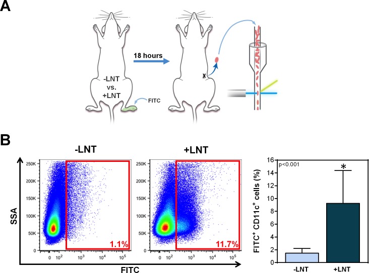
Fig 5. LNT increases migration of DCs to inguinal lymph node.
A) Schematic diagram of the experimental protocol. Ten weeks after surgery with or without LNT, mice underwent FITC painting of the ipsilateral distal hindlimb. Eighteen hours later, the number of DCs migrating to the inguinal lymph node was analyzed using flow cytometry. B) Left panel: Representative flow cytometry plots of inguinal lymph node cells gated for side scatter analysis (SSA) and FITC. The red boxes indicate the gating for FITC+ DCs. Right panel: Quantification of FITC+ DCs in the inguinal lymph nodes of mice treated with or without LNT.

