Figure 7.
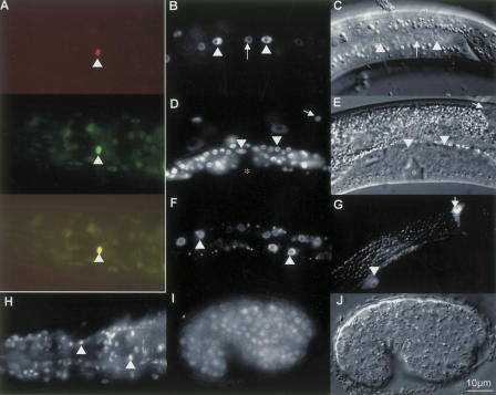
DIN-1 proteins are nuclear localized. (A–F) din-1S::gfp. (G) din-1Lp::gfp. (H–J) din-1LBcDNA::gfp. (A, top) rfp fusion to sdf-9/phosphatase marks XXX cell in L1 (Ohkura et al. 2003). (Middle) din-1S::gfp is highly expressed in XXX. (Bottom) Merge of both. (B) L3, din-1S::gfp in seam (arrowhead), hypodermal cells (arrow). (C) DIC image. (D) L4, distal tip (arrow), somatic gonadal cells (arrowhead). (E) DIC image. Asterisk, vulva. (F) L2, intestinal cells. (G) L4, din-1L promoter construct in seam (arrowhead) and distal tip cell (arrow). (H) Adult, din-1LBcDNA::gfp in pharynx and head neurons. (I) embryo. (J) DIC image. Bar, 10 μm.

