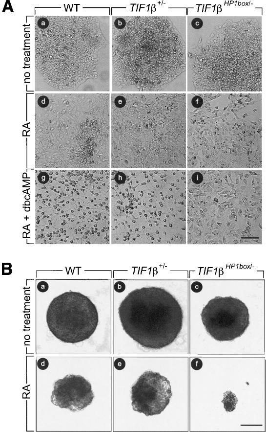
Figure 2.
TIF1βHP1box/- F9 cells differentiate into PrE cells but not into PE or VE cells. At day 0, 104 wild-type (WT), TIF1β+/-, and TIF1βHP1box/- cells were plated, and they were treated as described at day 1. (A) Wild-type (WT, panels a,d,g), TIF1β+/- (panels b,e,h), and TIF1βHP1box/- (panels c,f,i) cells were cultured either with vehicle (no treatment; panels a,b,c), in the presence of 1 μM tRA (panels d,e,f), or in the presence of 1 μM tRA + 250 μM dbcAMP (panels g,h,i). (B) Wild-type (WT, panels a,d), TIF1β+/- (panels b,e), and TIF1βHP1box/- (panels c,f) cells were cultured in bacterial Petri dishes with vehicle (no treatment; panels a–c) or in the presence of 50 nM RA (panels d–f) Cells were photographed at day 6 under a phase contrast microscope. Bar, 100 μm.

