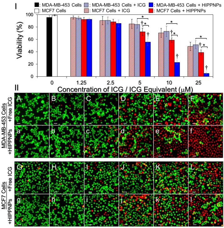
Fig 7. Phototoxicity of free ICG and HIPPNPs to HER2(+) and HER(-) breast cancer cells in vitro.
(I) Viabilities of MDA-MB-453 and MCF7 cells pre-treated with freely dissolved ICG or HIPPNPs in different doses and subjected to light illumination afterward. Both free ICG and HIPPNPs were exploited in concentrations of 0 (without agents; blank control), 1.25, 2.5, 5, 10, and 25 μM ICG equivalent for each type of cells and were co-cultured with the cells for only 4 h. The light illumination was performed by using a 808-nm CW laser with intensity of 6 W/cm2 for 5 min after the free ICG or HIPPNPs were removed. The cellular viability was determined by hemocytometry with trypan blue exclusion method immediately after the laser treatment. Values are mean ± SD (n = 3). *P < 0.05. †P < 0.05 as compared to the blank control. (II) Representative photomicrographic images of calcein-AM/PI-stained MDA-MB-453 (A/a–F/f) or MCF7 (G/g–L/l) cells after NIR laser irradiation. Before NIR illumination, cells were treated with free ICG (A—L) or HIPPNPs (a—l) in 0 (A/a and G/g), 1.25 (B/b and H/h), 2.5 (C/c and I/i), 5 (D/d and J/j), 10 (E/e and K/k), and 25 (F/f and L/l) μM ICG equivalent for 4 h and followed by PBS wash. The green cells stained by calcein-AM and the red cells stained by PI represent live and dead cells, respectively. All stained cells were suspended in PBS and the images were photographed by using a fluorescent microscope at 200X magnification. Scale bar = 50 μm.

