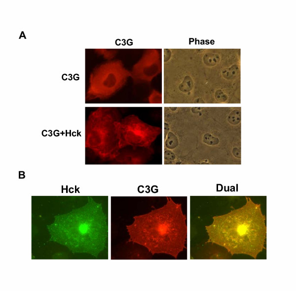
Figure 1.
Subcellular localization of C3G. (A) Cos-1 cells grown on coverslip were either transfected with C3G or cotransfected with Hck and indirect immunofluorescence staining performed using anti-C3G antibodies and Cy3 conjugated anti rabbit secondaries. (B) Cells transfected with Hck and C3G were stained for both the antigens as described in Materials and Methods. Hck was visualized using FITC conjugated secondaries and C3G by Cy3 conjugated secondaries. The dual panel shows the merged image of an optical section taken using the confocal microscope where the yellow signal generated shows colocalization of the two proteins.

