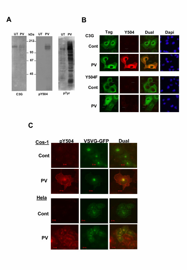
Figure 5.
Phosphorylation of endogenous C3G upon activation of endogenous tyrosine kinases. (A) Cells were either left untreated (UT) or treated with pervanadate (PV) and western blotting was performed using pY504 antibody. The same blot was reprobed with C3G to show the presence of endogenous C3G in these cells. (B) Cos-1 cells on coverslips were transfected with either C3G or Y504F mutant of C3G and fixed without any treatment (cont.) or after pervanadate treatment (PV). Dual labeling was performed using the tag antibodies (stained with FITC) and pY504 antibody (stained with Cy3). Panels show optical sections obtained by confocal microscopy. (C) Cos-1 and HeLa cells grown on coverslips and transfected with VSVG-GFP were fixed without any treatment (control) or after treatment with pervanadate and stained for pY504 expression. GFP fluorescence was used to visualize the staining pattern of VSVG-GFP protein. Optical sections taken using the apotome are shown. Areas of colocalization are seen from the yellow color generated in the merged images.

