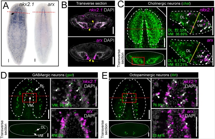
Figure 1. nkx2.1 and arx are expressed by VM neural cell types.
(A) WISH images of nkx2.1 and arx expression. Black arrow highlights CNS expression. (B) Single confocal plane from transverse sections of FISH images depicting expression of nkx2.1 and arx within the CNS. Yellow arrows highlight nkx2.1 and arx expression within stem cell-rich area. (C–E) For each set of images, the upper left panels are projections of confocal images from FISH experiments showing all cholinergic, GABAergic, or octopaminergic neurons in wild-type animals. Solid red boxes represent region of interest for the right panels, which are single confocal planes from dFISH experiments, showing co-expression of nkx2.1 or arx within a given neuron subtype. Solid yellow lines in (C) demarcate the division line between the VM and DL brain regions. White arrows in dFISH panels indicate double-positive cells. Solid white lines represent the border of the animal. Dashed white lines represent the approximate border of the CNS. Red dashed lines indicate approximate plane for transverse sections. Scale bar = 100 µm. CNS, Central nervous system; DL, Dorsal-lateral; FISH, Fluorescent in situ hybridization; VM, Ventral-medial.


