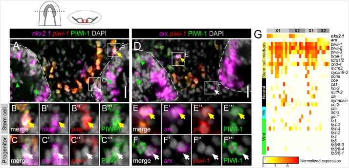
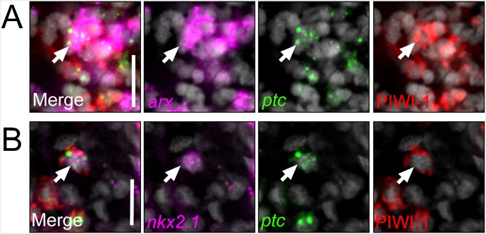
Figure 5. nkx2.1 and arx are expressed in stem cells and post-mitotic progenitors.
(A–F) Single confocal planes of dFISH images combined with immunolabelling, displaying expression of nkx2.1 and arx within stem cells (yellow arrows) and post-mitotic progenitors (white arrows). Green arrowheads highlight post-mitotic progenitors within the brain lobes. Boxed cells are enlarged below and individual channels are split. Dashed lines mark the outer border of the brain lobes. Cartoon depicts region of interest for all confocal images. Scale bars = 20 µm. (G) Heatmap depicting normalized expression of stem cell markers, genes associated with neurons, hh, and Wnt transduction genes, within 31 individually sequenced head stem cells (X1) or stem cells + progeny (X2) that also express arx or nkx2.1.


