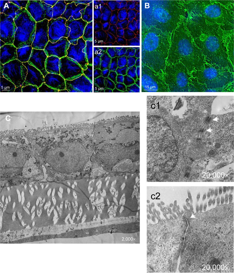
Figure 1.
Immunofluorescence and TEM of the Caco/2/ISO-HAS-1 coculture.
Notes: ([A], a1 and a2) Immunofluorescence staining of tight junctional proteins (ZO-1, red signal [A], a1) and adherens junctional proteins (beta-catenin, green signal [A], a2) of Caco-2 after 21 days in coculture with ISO-HAS-1 on the opposite side of the filter membrane. ZO-1 and beta-catenin appeared well developed. TEM images corroborate these observations, desmosomal ([C] c1, arrows) as well as tight junctional ([C] c2, arrows) complexes appear well defined. (B) CD31 staining of the ISO-HAS-1 cells underneath the Caco-2 layer. A well-developed endothelial monolayer could be detected. Nuclei (blue signal, Hoechst 33342) scale bar for fluorescence images (A), a1 and a2: 5 μm; (B) 15 μm. (C) TEM cross-section through a transwell filter membrane with Caco-2 on the top side and ISO-HAS-1 on the bottom side (magnifications C: 2,000×; c1, c2: 20,000×).
Abbreviations: TEM, transmission electron microscopy; ZO-1, zona occludens-1.

