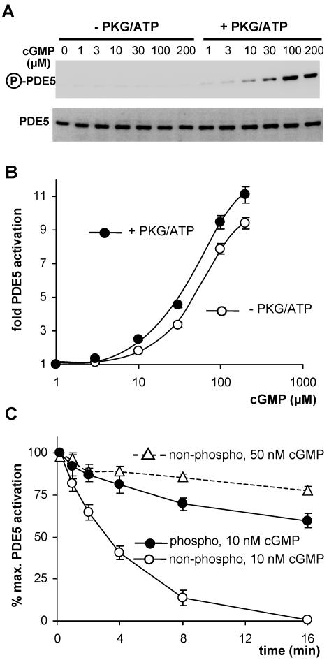
Figure 7.
Effect of phosphorylation on activation and deactivation of PDE5. (A) Western blot detection of phosphorylated PDE5 (top) after incubation of cytosols with increasing cGMP concentrations in the absence of presence of PKG/ATP; bottom shows loading of PDE5. (B) Activation of PDE5 by preincubation of cytosols with increasing cGMP concentrations. (C) Deactivation of PDE5. PDE5 was activated by preincubation with 30 μM cGMP in the presence or absence of PKG/ATP to obtain phospho- or a nonphospho-PDE5. Then samples were diluted to yield a cGMP concentration of 10 nM (solid lines) and further incubated. At the indicated time points, PDE5 activity was determined. The dashed line represents deactivation of nonphospho-PDE5 at a cGMP concentration of 50 nM.

