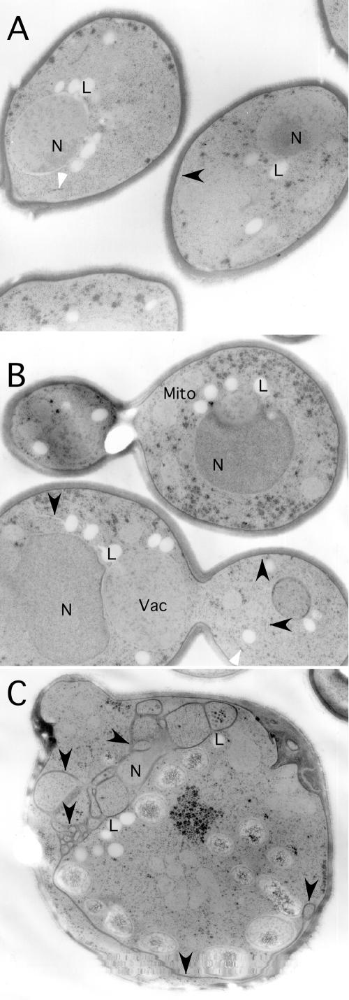
Figure 5.
Glo3-R59K causes accumulation of ER membrane. glo3Δ cells carrying the MET3pr-GLO3 plasmid (A), an “empty” vector plasmid (B), or the MET3pr-glo3-R59K plasmid (C) were shifted to medium lacking methionine and grown for 6 h at 30°C. Cells were collected and fixed for examination by electron microscopy. N, nucleus; Mito, mitochondria; and Vac, vacuole. The black arrow-heads point to peripheral ER in all three panels. The white arrow-heads indicate Golgi cisternae. Lipid bodies (L) are present in all strains due to growth in minimal media.

