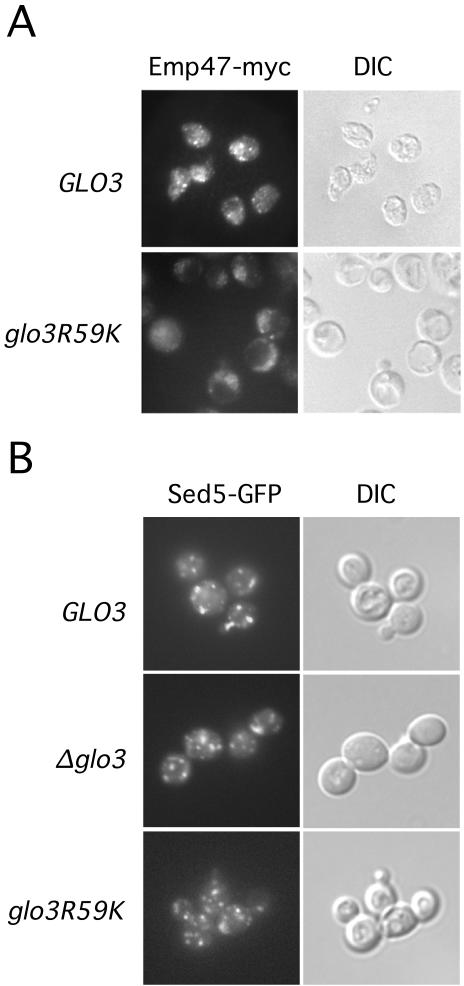
Figure 7.
Retrograde cargo localization requires intact Glo3 function. (A) Cells harboring the MET3pr-GLO3 or MET3pr-glo3-R59K genes were transferred to medium lacking methionine to induce gene expression. After 6 h, cells were processed for fluorescence microscopy as described previously. The retrograde cargo protein Emp47-myc was detected by Alexa 488 fluorescence (left). Cell morphology (differential interference contrast) is displayed in panels on the right. (B) Sed5-GFP is localized to the Golgi. glo3Δ cells carrying the MET3pr-GLO3 plasmid, an empty vector plasmid or the MET3pr-glo3-R59K plasmid and plasmid expressing Sed5-GFP were shifted to medium lacking methionine and grown for 6 h at 30°C. Cells were immediately examined for Sed5-GFP localization.

