Figure 3.
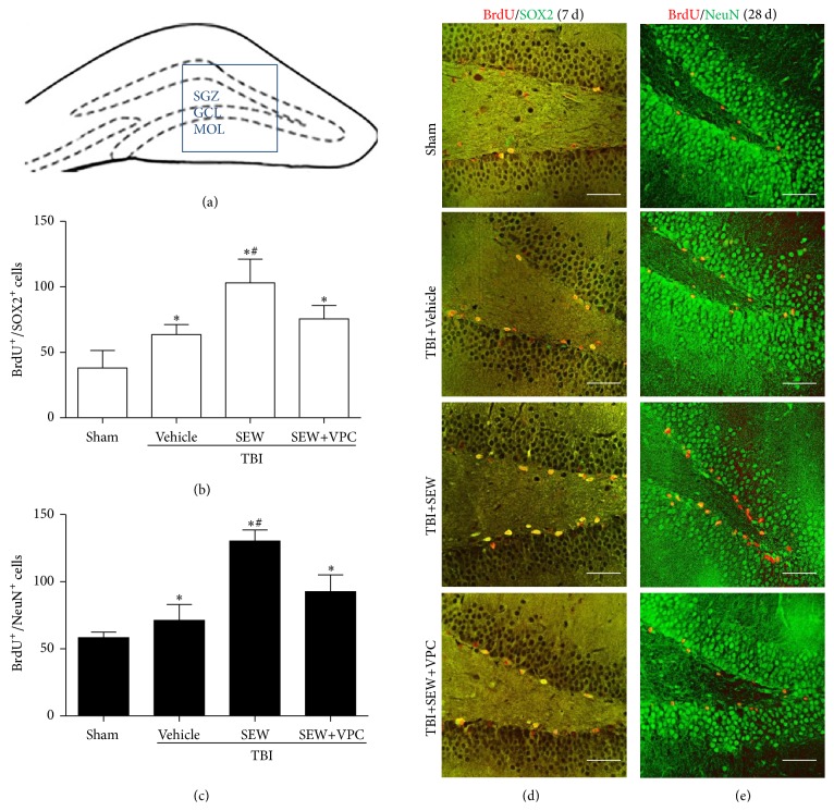
S1PR1 activation enhanced NSCs proliferation and neuronal differentiation in hippocampal dentate gyrus (DG) after TBI. (a) Coronal diagram of rat hippocampus, the subgranular zone, granular cells layer, and molecular layer of DG were, respectively, marked by SGZ, GCL, and MOL, the blue pane representing one of microscopic visual fields for counting immunolabeled cells of immunofluorescence (IF). (b) NSCs proliferation in SGZ at 7 days after TBI was assessed by BrdU/SOX2 double-labeling IF. Quantitation analysis showed that, relative to sham group (n = 3), brain injury induced more double-positive cells in TBI+Vehicle group (n = 6). Following the treatment of SEW2871, the number of BrdU+/SOX2+ cells further increased in TBI+SEW group (n = 6). Reversely, administration of VPC23019 resulted in a significant reduction of BrdU+/SOX2+ cells in TBI+SEW+VPC group (n = 6). (c) Neuronal differentiation of NSCs in SGZ at 28 days after TBI was assessed by BrdU/NeuN double-labeling IF. Statistical data indicated that BrdU+/NeuN+ cells increased in TBI+Vehicle group (n = 6) compared with sham group (n = 3). And the double-positive cells increased at even higher level after SEW2871 treatment in TBI+SEW group (n = 6). However, VPC23019 administration caused an evident decrease of BrdU+/NeuN+ cells in TBI+SEW+VPC group (n = 6). (d) and (e) Representative IF microphotographs of hippocampus immunolabeled with BrdU/SOX2 and BrdU/NeuN in all the above four groups at 7 days and 28 days after trauma. Scale bar: 50 μm. ∗ P < 0.05 versus sham group; # P < 0.05 versus TBI+Vehicle group or TBI+SEW+VPC group.

