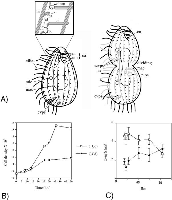
Figure 1.
(A) Schematic representation of MT-based organelles in Tetrahymena. The intracytoplasmic, macronuclear, oral fiber, and cytoproct MTs are not shown. An interphase (left) and dividing (right) cell are shown. The inset shows a magnification of a region of the cortex surrounding two adjacent BBs. In the dividing cell, we distinguish between preformed (black) and newly made organelles (gray). We also use gray to depict preformed organelles with a relatively high rate of subunit exchange, even though some of them are maintaining structural integrity (LMs and CVPs). Note the presence of an anterior and posterior invariant zone in the dividing cell. ss, separation spindle (anaphase B) of the micronucleus; mic, micronucleus; mac, macronucleus; cvps, contractile vacuole pores; ncvps, new contractile vacuole pores, m, oral membranelles; um, undulating membrane; oa, oral apparatus; noa, new oral apparatus; lm, longitudinal MT; tm, transverse MT; kd, kinetodesmal fiber (nonmicrotubular); pc, postciliary MT; bb, basal body. (B) Growth of βDDDE440-CD cells in the presence (open squares) and absence of Cd (solid squares). The mutant cells undergo only two to three complete cell divisions before undergoing a cytokinesis arrest. (C) Length of old (rectangles) and new cilia (filled circles) in the βDDDE440-CD cells as a function of time of incubation in the medium lacking Cd. Data points represent average length of cilia (±SD), calculated for 20-50 cilia per each time point.

