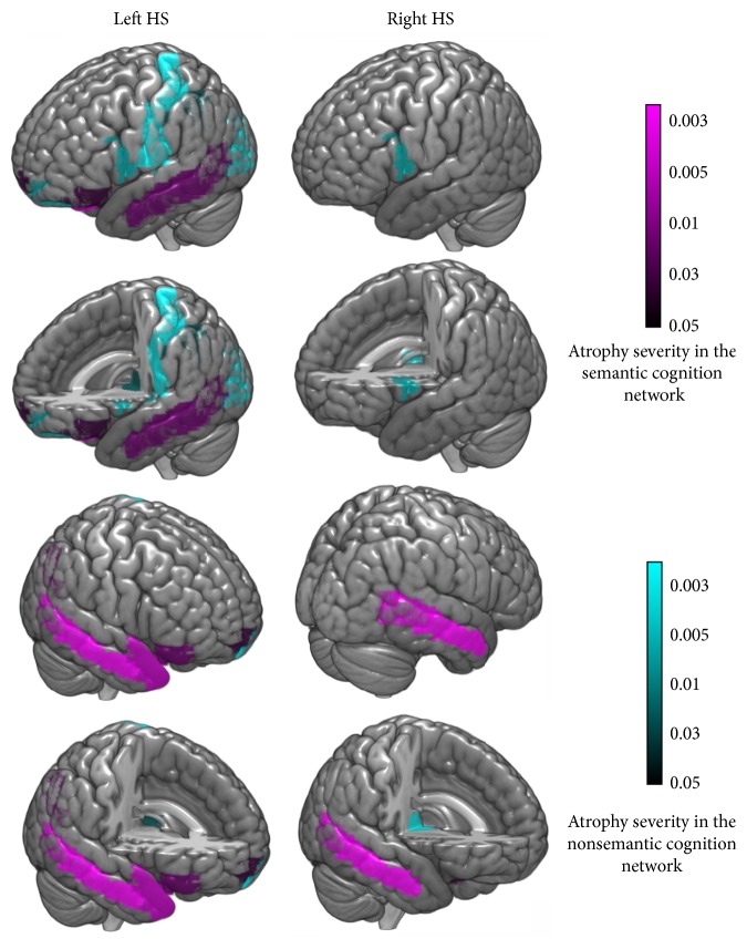
Figure 1.
Gray matter volume (GMV) loss of patients with left HS or right HS (FWE corrected, p = 0.05; minimum cluster size 10). In the left HS subgroup, GMV loss was observed in 12 ROIs in the ipsilateral lobe (including the IFG, vmPFC, hippocampus, and MTG in the semantic cognition network) and 7 ROIs in the contralateral lobe (including the IFG, vmPFC, parahippocampus, fusiform, AG, tpSTG, tpMTG, and MTG, all of which were components of the semantic cognition network); in the right HS subgroup, GMV loss was seen in 2 ROIs in the ipsilateral lobe and the MTG in the contralateral lobe.

