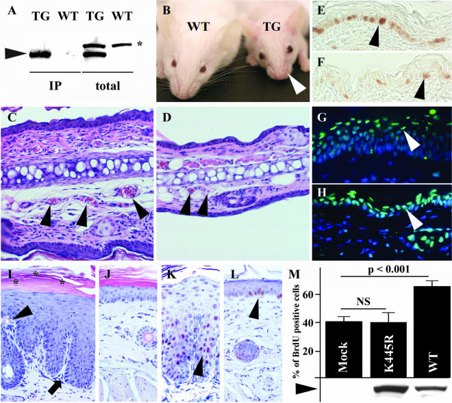
Figure 2.
Comparison of K14-Bmx transgenic and control littermate mice. (A) Bmx expression (arrowhead) in TG mice analyzed by Western blotting of immunoprecipitates (IP) or total skin lysates (total). The asterisk indicates a nonspecific polypeptide. (B) Note the absent whiskers in TG mouse. H&E-stained cross sections from the ears of TG and WT mice (C and D). The arrowheads point to blood vessels in the cross sections. PCNA staining of the epidermis of back skin in TG (E) and WT mice (F). The arrowheads point to the proliferating basal cells. Sections of TG (G) and WT (H) ears stained for apoptotic cells (TUNEL; green, arrowheads) and cell nuclei (Hoechst; blue). Sections of tail skin of TG (I) and WT mice (J). Arrowhead shows the dermal papillae containing blood capillaries; arrow indicates the rete ridge-like changes in the skin; asterisk mark parakeratosis (retained cell nuclei in the horny layer of the epidermis). PCNA staining of tail sections of TG (K) and WT mice (L). Arrowheads indicate PCNA positive nuclei in the skin. (M) The percentage of BrdU-positive cells in MMC-E epithelial cells expressing mock vector (Mock), kinase-deficient Bmx (K445R) or Bmx (WT). Bars represent average counts from four separate infections and below are shown the relative protein amounts as detected by Western blotting. NS, not significant.

