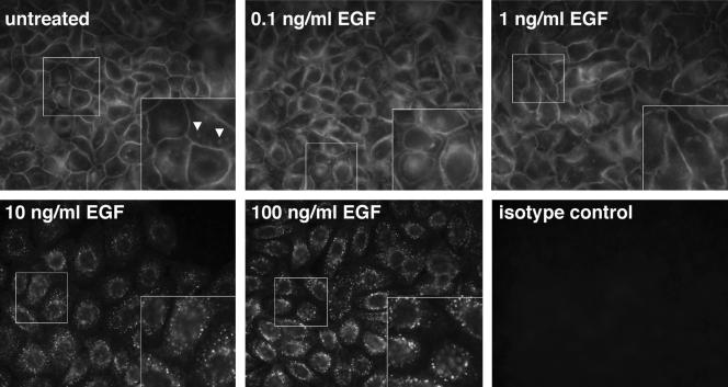
Figure 6.
Localization of ErbB1 receptors in response to different concentrations of EGF. NHKs were deprived of GFs for 48 h and then incubated for 20 min in fresh basal M154 medium. At that point, they were either left untreated or stimulated with 0.1, 1, 10, and 100 ng/ml EGF for 10 min at 37°C, as indicated on each panel. ErbB1 receptor localization was then assessed by immunofluorescence by using a mixture of three mAbs recognizing the extracellular domain of ErbB1. Insets show 2× magnifications of the segment of each image enclosed in the smaller white box. Arrowheads indicate the patchy ErbB1 staining referred to in the text. The results shown are from a single experiment, and are representative of three independent experiments yielding similar results.

