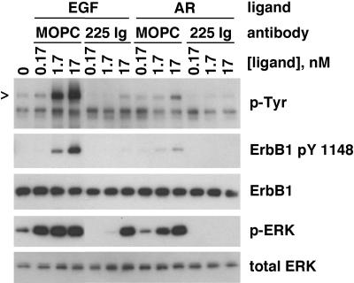
Figure 9.
Stimulation of ERK phosphorylation by AR. NHKs were depleted of GFs in basal M154 medium for 48 h and then incubated for 20 min with fresh basal M154 containing 5 μg/ml 225 IgG or 5 μg/ml MOPC 21. Then, the indicated concentrations of AR or EGF were added, and incubation was continued for another 10 min. PBS was added rather than AR or EGF in the lane labeled 0. Nonionic detergent lysates were then prepared and subjected to western blotting (20 μg protein/lane). Replicate blots were prepared and decorated with the antibodies indicated to the right of the autoradiographs. Arrowhead indicates mobility of ErbB1. For comparison with other figures, 0.17 nM EGF is 1 ng/ml. Note the decreased potency and effect of rhAR, compared with rhEGF.

