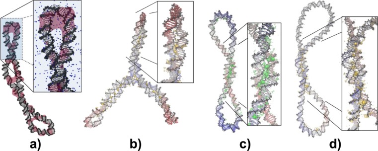
Fig. 2.
Regions of high positive counterion density around negatively supercoiled DNA minicircles. a Structure from explicitly solvated atomistic MD of a negatively supercoiled 336-bp minicircle (σ ≈ −0.1) showing regions highly populated by counterions over 20 ns as pink isosurfaces. b–d Averaged structure of a 339-bp minicircle solvated in 100 mM Ca(Cl)2 (highly negatively supercoiled; σ ≈ −0.2) (b), of a 260-bp minicircle in 200 mM NaCl (σ ≈ −0.08) (c) and of a 339-bp minicircle in 100 mM Ca(Cl)2 (σ ≈ −0.07) (d) obtained from a superposition of 1000 snapshots corresponding to the last 10 ns of 100-ns MD trajectories. Ca2+ density peaks are shown in yellow and Na+ peaks in green

