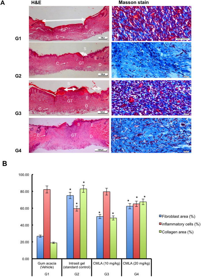
Figure 4. Effect of CMLA on histological section (H&E) and Masson’s Trichrome staining of healed wound on day 15 post-surgery.
The arrow showed epithelialization. S-Scab; E-Epidermis; D-Dermis; GT-Granulation tissue; C-Capillaries. (G1) Gum acacia group showing incomplete wound healing enclosure and few degree and alignment of collagen (G2) Intrasit gel group showing complete wound healing enclosure and a greater degree and alignment of collagen (G3) 10 mg/ml of CMLA group showing narrow scar region of wound closure and moderate degree and alignment of collagen. (G4) 20 mg/ml of CMLA group showing complete wound healing enclosure and more degree and alignment of collagen (H&E stain 4×), (Masson’s Trichrome stain 100×). Image analysis was executed using an optical image analyzer (ImagePro Plus 4.5, Media Cybernetics, Silver Spring, MD). Data was expressed as the mean ±SEM (n = 6) and analyzed using one way analysis of variance ANOVA followed by Dunnett’s post hoc test for average comparison on SPSS 18.0. Mean values ± SEM were used. Significance was defined as *p < 0.05 compared to G1.

