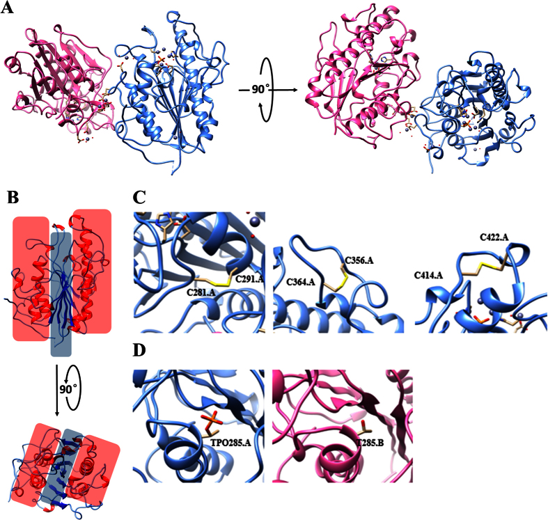
Figure 1. Overall structure of MCR-1-ED.
(A) Front and top view of the crystal structure of MCR-1-ED. Two molecules in one asymmetric unit are labeled with cornflower blue and hot pink for chain A and B respectively. (B) Front and top view of the “sandwich” conformation of MCR-1-ED covered by red (α-helix layer) and blue (β-sheet layer) shadows. Secondary structures of α-helixes and β-stands are colored in red and blue respectively. (C) Three pairs of disulfide bonds of MCR-1-ED labeled with yellow on chain A. The other three are located at the same positions on chain B. (D) Threonine 285 of MCR-1-ED with and without phosphorylation.

